New TCIA Dataset Page

[vc_row type=”in_container” full_screen_row_position=”middle” column_margin=”default” column_direction=”default” column_direction_tablet=”default” column_direction_phone=”default” scene_position=”center” top_padding=”20″ text_color=”dark”…

Collection Manager REST API Page

[vc_row type=”in_container” full_screen_row_position=”middle” column_margin=”default” column_direction=”default” column_direction_tablet=”default” column_direction_phone=”default” scene_position=”center” text_color=”dark” text_align=”left”…

DICOM-LIDC-IDRI-NODULES

DOI: 10.7937/TCIA.2018.h7umfurq | Analysis Result

This dataset contains standardized DICOM representation of the annotations and characterizations collected by the LIDC/IDRI initiative, originally stored in XML and available in the TCIA LIDC-IDRIย collection . Only the nodules that were deemed to be greater or equal to 3 mm in the largest planar dimensions have been annotated and characterized by the...

Research Activities Page

[vc_row type=”in_container” full_screen_row_position=”middle” column_margin=”default” column_direction=”default” column_direction_tablet=”default” column_direction_phone=”default” scene_position=”center” text_color=”dark” text_align=”left”…

Analyses of Existing TCIA Datasets Page

[vc_row type=”in_container” full_screen_row_position=”middle” column_margin=”default” column_direction=”default” column_direction_tablet=”default” column_direction_phone=”default” scene_position=”center” top_padding=”40″ text_color=”dark”…

DICOM-SR-BREAST-CLINICAL

DOI: 10.7937/TCIA.2019.wgllssg1 | Analysis Result

The Data Integration & Imaging Informatics (DI-Cubed) project explored the issue of lack of standardized data capture at the point of data creation, as reflected in the non-image data accompanying various TCIA breast cancer collections. The work addressed the desire for semantic interoperability between various NCI initiatives by aligning on common clinical metadata elements and supporting use cases that connect clinical,...

BRATS-TCGA-LGG

DOI: 10.7937/K9/TCIA.2017.GJQ7R0EF | Analysis Result

This data container describes both computer-aided and manually-corrected segmentation labels for the pre-operative multi-institutional scans of The Cancer Genome Atlas (TCGA) Low Grade Glioma (LGG) collection, publicly available in The Cancer Imaging Archive (TCIA), coupled with a rich panel of radiomic features along with their corresponding skull-stripped and...

[…] Robert, C. (2022). AutoComBat: a generic method for harmonizing MRI-based […]

RADIOMICS-TUMOR-PHENOTYPES

DOI: 10.7937/K9/TCIA.2014..UA0JGPDG | Analysis Result

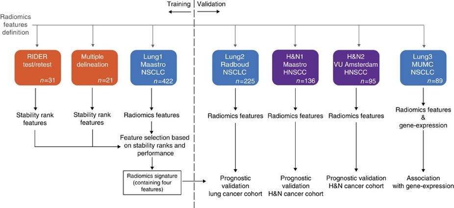
This data applies a radiomic approach to computed tomography data of 1,019 patients with lung or head-and-neck cancer which are described in Nature Communications (http://doi.org/10.1038/ncomms5006).ย  The various arms of the study are represented in TCIA as distinct Collections includingย NSCLC-Radiomicsย (Lung1),ย 

RSNA-ASNR-MICCAI-BRATS-2021

DOI: 10.7937/jc8x-9874 | Analysis Result

This dataset includes brain MRI scans of adult brain glioma patients, comprising of 4 structural modalities (i.e., T1, T1c, T2, T2-FLAIR) and associated manually generated ground truth labels for each tumor sub-region (enhancement, necrosis, edema), as well as their MGMT promoter methylation status. These scans are a collection of data from existing TCIA collections, but also cases provided by individual institutions...