Meningioma-SEG-CLASS | Segmentation and Classification of Grade I and II Meningiomas from Magnetic Resonance Imaging: An Open Annotated Dataset
DOI: 10.7937/0TKV-1A36 | Data Citation Required | 1.1k Views | 4 Citations | Image Collection
| Location | Species | Subjects | Data Types | Cancer Types | Size | Status | Updated | |
|---|---|---|---|---|---|---|---|---|
| Brain and Spinal Cord | Human | 96 | MR, RTSTRUCT, Demographic, Diagnosis, Measurement | Meningioma | Clinical | Limited, Complete | 2023/02/13 |
Summary
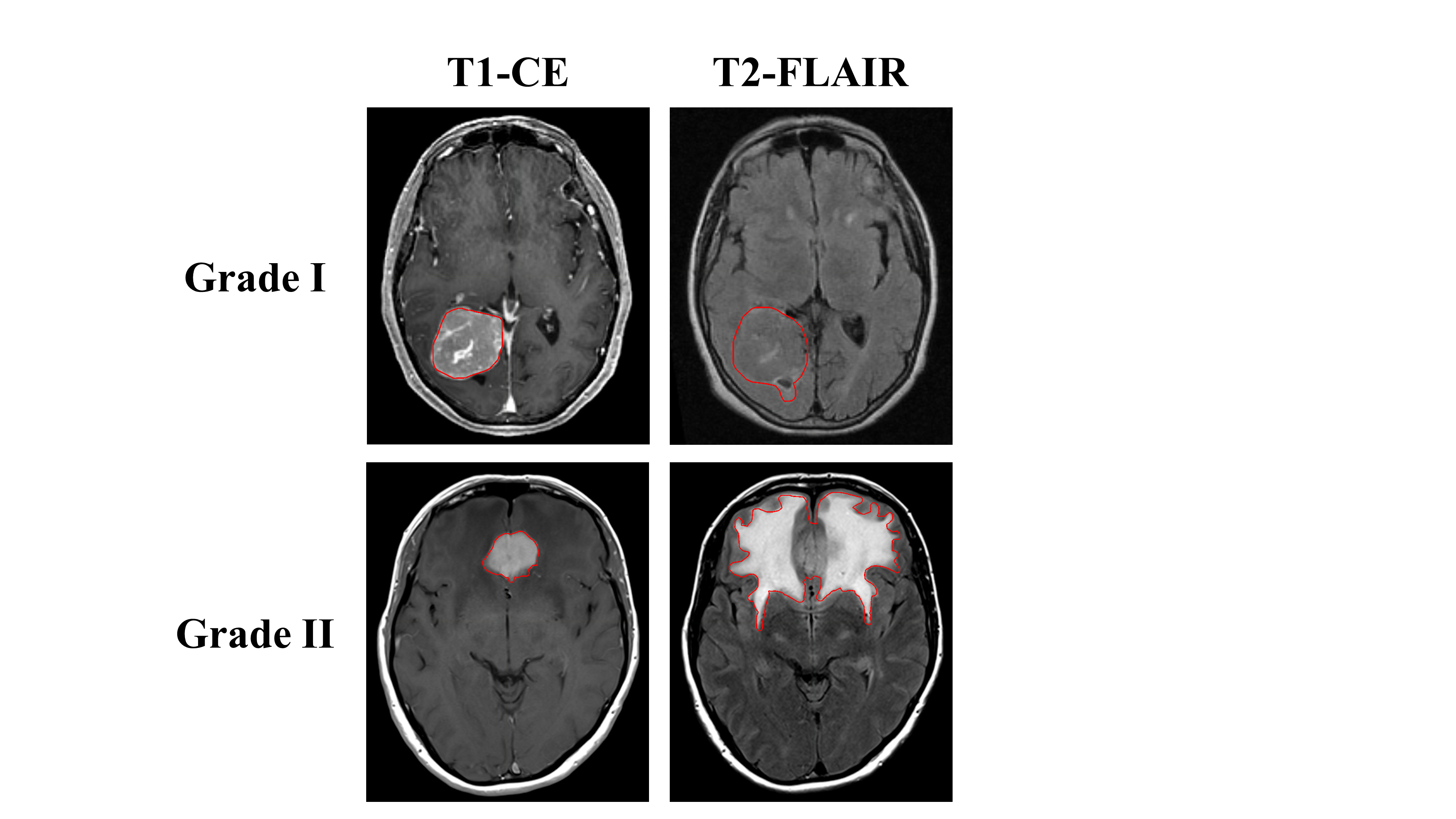
The study included 96 consecutive treatment naïve patients with intracranial meningiomas treated with surgical resection from 2010 to 2019. All patients had pre-operative T1, T1-CE, and T2-FLAIR MR images with subsequent subtotal or gross total resection of pathologically confirmed grade I or grade II meningiomas. A neuropathology team reviewed histopathology, including two subspecialty trained neuropathologists and one neuropathology fellow. The meningioma grade was confirmed based on current classification guidelines, most recently described in the 2016 WHO Bluebook. Clinical information includes grade, subtype, type of surgery, tumor location, and atypical features. Meningioma labels on T1-CE and T2-FLAIR images will also be provided in DICOM format. The hyperintense T1-contrast enhancing tumor and hyperintense T2-FLAIR and tumor were manually contoured on each MRI and reviewed by a central nervous system radiation oncologist specialist.
Data Access
Some data in this collection contains images that could potentially be used to reconstruct a human face. To safeguard the privacy of participants, users must sign and submit a TCIA Restricted License Agreement to help@cancerimagingarchive.net before accessing the data.
Version 1: Updated 2023/02/13
| Title | Data Type | Format | Access Points | Subjects | License | Metadata | |||
|---|---|---|---|---|---|---|---|---|---|
| Images and Radiation Therapy Structures | MR, RTSTRUCT | DICOM | Download requires NBIA Data Retriever |
96 | 180 | 674 | 47,520 | TCIA Restricted | View |
| Clinical data | Demographic, Diagnosis, Measurement | XLSX | CC BY 4.0 | — |
Citations & Data Usage Policy
Data Citation Required: Users must abide by the TCIA Data Usage Policy and Restrictions. Attribution must include the following citation, including the Digital Object Identifier:
Data Citation |
|
|
Vassantachart, A., Cao, Y., Shen, Z., Cheng, K., Gribble, M., Ye, J. C., Zada, G., Hurth, K., Mathew, A., Guzman, S., & Yang, W. (2023). Segmentation and Classification of Grade I and II Meningiomas from Magnetic Resonance Imaging: An Open Annotated Dataset (Meningioma-SEG-CLASS) (Version 1) [Data set]. The Cancer Imaging Archive. https://doi.org/10.7937/0TKV-1A36 |
Related Publications
Publications by the Dataset Authors
The authors recommended the following as the best source of additional information about this dataset:
Publication Citation |
|
|
Vassantachart, A., Cao, Y., Gribble, M., Guzman, S., Ye, J. C., Hurth, K., Mathew, A., Zada, G., Fan, Z., Chang, E. L., & Yang, W. (2022). Automatic differentiation of Grade I and II meningiomas on magnetic resonance image using an asymmetric convolutional neural network. In Scientific Reports (Vol. 12, Issue 1). Springer Science and Business Media LLC. https://doi.org/10.1038/s41598-022-07859-0 |
No other publications were recommended by dataset authors.
Research Community Publications
TCIA maintains a list of publications that leveraged this dataset. If you have a manuscript you’d like to add please contact TCIA’s Helpdesk.
