ReMIND | The Brain Resection Multimodal Imaging Database
DOI: 10.7937/3RAG-D070 | Data Citation Required | 3.6k Views | Image Collection
| Location | Species | Subjects | Data Types | Cancer Types | Size | Status | Updated | |
|---|---|---|---|---|---|---|---|---|
| Brain | Human | 114 | MR, SEG, US, Segmentation, Demographic, Histopathology, Molecular Test, Follow-Up, Classification | Brain Cancer | Clinical | Public, Complete | 2023/09/26 |
Summary
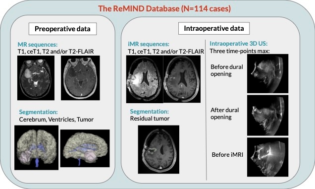
The Brain Resection Multimodal Imaging Database (ReMIND) contains pre- and intra-operative data collected on 114 consecutive patients who were surgically treated with image-guided tumor resection between 2018 and 2022. For all patients, preoperative MRI, 3D intraoperative ultrasound series, and intraoperative MRI are available. Additionally, each case typically contains segmentations, including the preoperative tumor, the pre-resection cerebrum, the previous resection cavity derived from the preoperative MRI (if applicable), and any residual tumor identified on the intraoperative MRI. In total, this collection contains 369 preoperative MRI series, 320 3D intraoperative ultrasound series, 301 intraoperative MRI series, and 356 segmentations. We expect this data to be a resource for computational research in brain shift and image analysis as well as for neurosurgical training in the interpretation of intraoperative ultrasound and intraoperative MRI. Preoperative MRI comprises four structural MRI sequences: native T1-weighted (T1), contrast-enhanced T1-weighted (ceT1), native T2-weighted (T2), and T2-weighted fluid-attenuated inversion recovery (T2-FLAIR). These scans were acquired before surgery using various scanners at multiple institutions, making their acquisition parameters heterogeneous. Unlike preoperative MRI, all intraoperative MRI were acquired using a 3T wide-bore (70 cm) MRI scanner. All iUS series were acquired using a tracked 2D neuro-cranial curvilinear transducer. The transducer was swept unidirectionally through the craniotomy at a slow, consistent speed. This specific motion, in conjunction with the tracking, enabled the reconstruction of a 3D volume from the tracked 2D sweeps. Various segmentations were created to assist the surgical resection. These include manual segmentations of the preoperative whole tumor, preoperative tumor target (i.e., the radiologically identifiable tumor specifically targeted for resection), resection cavity resulting from prior surgery (i.e., in case of reoperation), intraoperative residual tumor, and the automatic segmentations of cerebrum and ventricles (Brainlab AG, Munich, Germany). Only structures deemed necessary for the surgical resection by the attending neurosurgeon were segmented. Specifically, segmentations of the manual preoperative whole tumor (113 cases), preoperative tumor target segmentations (3 cases), manual previous resection cavity segmentations (21 cases), residual tumor segmentations (58 cases), and automated segmentations of the cerebrum (89 cases) and ventricles (54 cases). All cerebrum, ventricle, and tumor segmentations were created preoperatively during the surgical planning stage. In contrast, residual tumor segmentations were created intraoperatively from iMRI. Demographic information, including age, sex, and ethnicity, was obtained from the corresponding patient medical records. The age range of the included population was 20–76. The ratio of male:female was equal to 61:53. Moreover, clinico-pathologic data such as the tumor type, tumor grade, radiological characteristics upon contrast administration, tumor location, and the reoperation status were assessed by the treating neurosurgeons. Tumor type and grade were specified according to the World Health Organization (WHO) 2021 Classification of Tumors of the Central Nervous System. Additionally, tumors were classified into one of 3 categories based on proximity to the functional cortex (non-eloquent, near eloquent, and eloquent). The MRI and ultrasound images are provided in DICOM format. Segmentation files are provided in NRRD format (original format) and DICOM SEG (converted from NRRD). Data was converted from NRRD format to DICOM format using 3D Slicer (MR data), dicom3tools software (iUS data) and dcmqi (segmentation data). All MRI images were defaced using automatic affine registration or manual landmark registration with NiftyReg and the template and face mask provided in pydeface. The code of the algorithm is publicly available. Introduction:
Imaging data:
Segmentation data:
Clinical metadata:
Pre-processing:
Data Access
Version 1: Updated 2023/09/26
| Title | Data Type | Format | Access Points | Subjects | License | Metadata | |||
|---|---|---|---|---|---|---|---|---|---|
| Images and Segmentations | MR, SEG, US | DICOM | Download requires NBIA Data Retriever |
114 | 228 | 1,346 | 85,733 | CC BY 4.0 | View |
| Segmentations | Segmentation | NRRD | Download requires IBM-Aspera-Connect plugin |
113 | 0 | 113 | CC BY 4.0 | — | |
| Clinical data | Demographic, Histopathology, Molecular Test, Follow-Up, Classification | CSV | CC BY 4.0 | — |
Additional Resources for this Dataset
The following external resources have been made available by the data submitters. These are not hosted or supported by TCIA, but may be useful to researchers utilizing this collection.
- ReMIND2Reg dataset – The goal of the ReMIND2Reg dataset is to address the registration problem of multi-parametric pre-operative MRI and intra-operative 3D ultrasound (iUS) images. Specifically, we focus on the challenging problem of pre-operative to post-resection registration, requiring the estimation of large deformations and tissue resections.
The NCI Cancer Research Data Commons (CRDC) provides access to additional data and a cloud-based data science infrastructure that connects data sets with analytics tools to allow users to share, integrate, analyze, and visualize cancer research data.
- Imaging Data Commons (IDC) (Imaging Data)
Citations & Data Usage Policy
Data Citation Required: Users must abide by the TCIA Data Usage Policy and Restrictions. Attribution must include the following citation, including the Digital Object Identifier:
Data Citation |
|
|
Juvekar, P., Dorent, R., Kögl, F., Torio, E., Barr, C., Rigolo, L., Galvin, C., Jowkar, N., Kazi, A., Haouchine, N., Cheema, H., Navab, N., Pieper, S., Wells, W. M., Bi, W. L., Golby, A., Frisken, S., & Kapur, T. (2023). The Brain Resection Multimodal Imaging Database (ReMIND) (Version 1) [dataset]. The Cancer Imaging Archive. https://doi.org/10.7937/3RAG-D070 |
Acknowledgements
We would like to acknowledge the individuals and institutions that have provided data for this collection:
NIH grants R01EB027134, 5P41EB015902, and P41EB028741
- We would also like to acknowledge the support and contribution of our collaborating neurosurgeons within the Department of Neurosurgery at Brigham and Women’s Hospital (Boston, USA).
Related Publications
Publications by the Dataset Authors
The authors recommended the following as the best source of additional information about this dataset:
Publication Citation |
|
|
Juvekar, P., Dorent, R., Kogl, F., Torio, E., Barr, C., Rigolo, L., Galvin, C., Jowkar, N., Kazi, A., Haouchine, N., Cheema, H., Navab, N., Pieper, S., Wells, W. M., Bi, W. L., Golby, A., Frisken, S., & Kapur, T. (2023). ReMIND: The Brain Resection Multimodal Imaging Database. Cold Spring Harbor Laboratory. https://doi.org/10.1101/2023.09.14.23295596 |
The Collection authors recommend these readings to give context to this dataset
- Juvekar, P., Dorent, R., Kögl, F., Torio, E., Barr, C., Rigolo, L., Galvin, C., Jowkar, N., Kazi, A., Haouchine, N., Cheema, H., Navab, N., Pieper, S., Wells, W. M., Bi, W. L., Golby, A., Frisken, S., & Kapur, T. (2024). ReMIND: The Brain Resection Multimodal Imaging Database. In Scientific Data (Vol. 11, Issue 1). Springer Science and Business Media LLC. https://doi.org/10.1038/s41597-024-03295-z
Research Community Publications
TCIA maintains a list of publications that leveraged this dataset. If you have a manuscript you’d like to add please contact TCIA’s Helpdesk.
