LDCT-AND-PROJECTION-DATA

By
DOI: 10.7937/9NPB-2637 | Image Collection

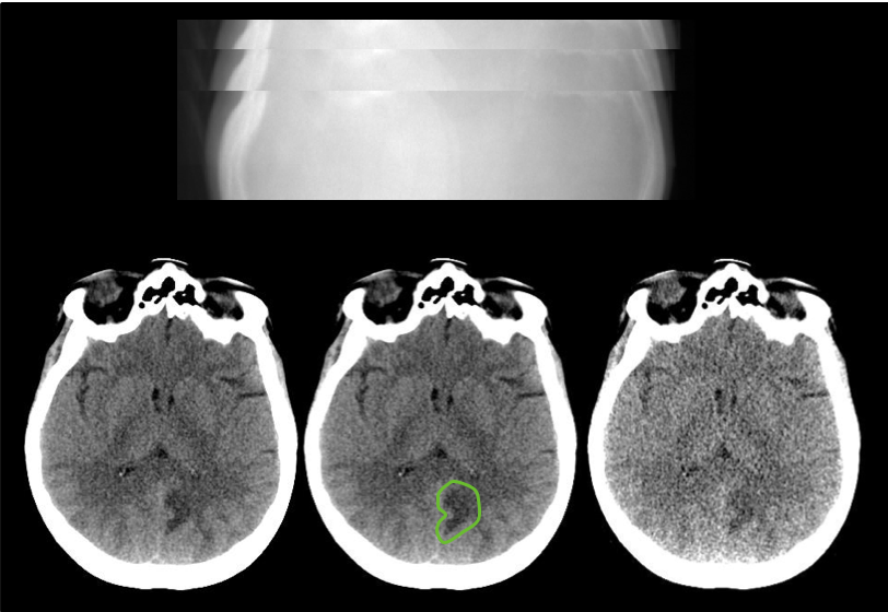
Investigators at the Mayo Clinic, with funding from the National Institute of Biomedical Imaging and Bioengineering (EB 017095 and EB 017185), have built a library of CT patient projection data in an open and vendor-neutral format. This format, referred to as DICOM-CT-PD (Additional information regarding the CT projection data format in the article by Chen et al at doi:

Read More

LGG-1P19QDELETION

By
DOI: 10.7937/K9/TCIA.2017.DWEHTZ9V | Image Collection

These MRIs are pre-operative examinations performed in 159 subjects with Low Grade Gliomas (WHO grade II & III). Segmentation of tumors in three axial slices that include the one with the largest tumor diameter and ones below and above are provided in NiFTI format.  Tumor grade and histologic type are also available.  All of these subjects have biopsy proven 1p/19q results, performed using FISH.  For the...

Read More

IVYGAP

By
DOI: 10.7937/K9/TCIA.2016.XLwaN6nL | Image Collection

This data collection consists of MRI/CT scan data for brain tumor patients that form the cohort for the resource Ivy Glioblastoma Atlas Project (Ivy GAP). There are 390 studies for 39 patients that include pre-surgery, post-surgery and follow up scans. The Ivy GAP is a collaborative partnership between the Ben and Catherine Ivy Foundation, which generously provided the financial...

Read More

ISPY2

By
DOI: 10.7937/TCIA.D8Z0-9T85 | Image Collection

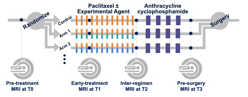
I-SPY 2 (Investigation of Serial studies to Predict Your Therapeutic Response with Imaging And moLecular analysis 2) is an ongoing, multi-center trial (NCT01042379) designed to quickly evaluate the efficacy of new agents for breast cancer in neoadjuvant chemotherapy (NAC) setting. Women aged ≥18 years diagnosed with locally advanced breast cancer (tumor...

Read More

PDMR-833975-119-R

By
DOI: 10.7937/TCIA.0ECK-C338 | Image Collection

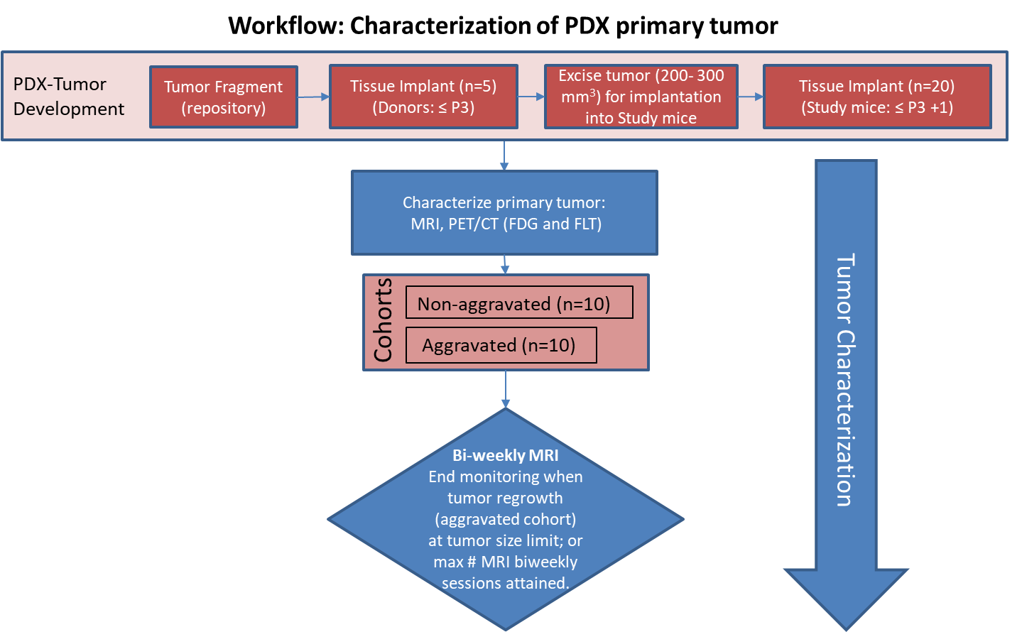
Characterization of tissue using in vivo non-invasive imaging is the foundation of Radiology and is clinically used for detection and measurement of disease burden in oncology.  With the migration of imaging to digital media the possibility for advanced mathematically based imaging biomarkers was realized. As part of this endeavor, researchers have developed various algorithms using neural networks, and classification...

Read More

PDMR-425362-245-T

By
DOI: 10.7937/TCIA.2020.7YRS-7J97 | Image Collection

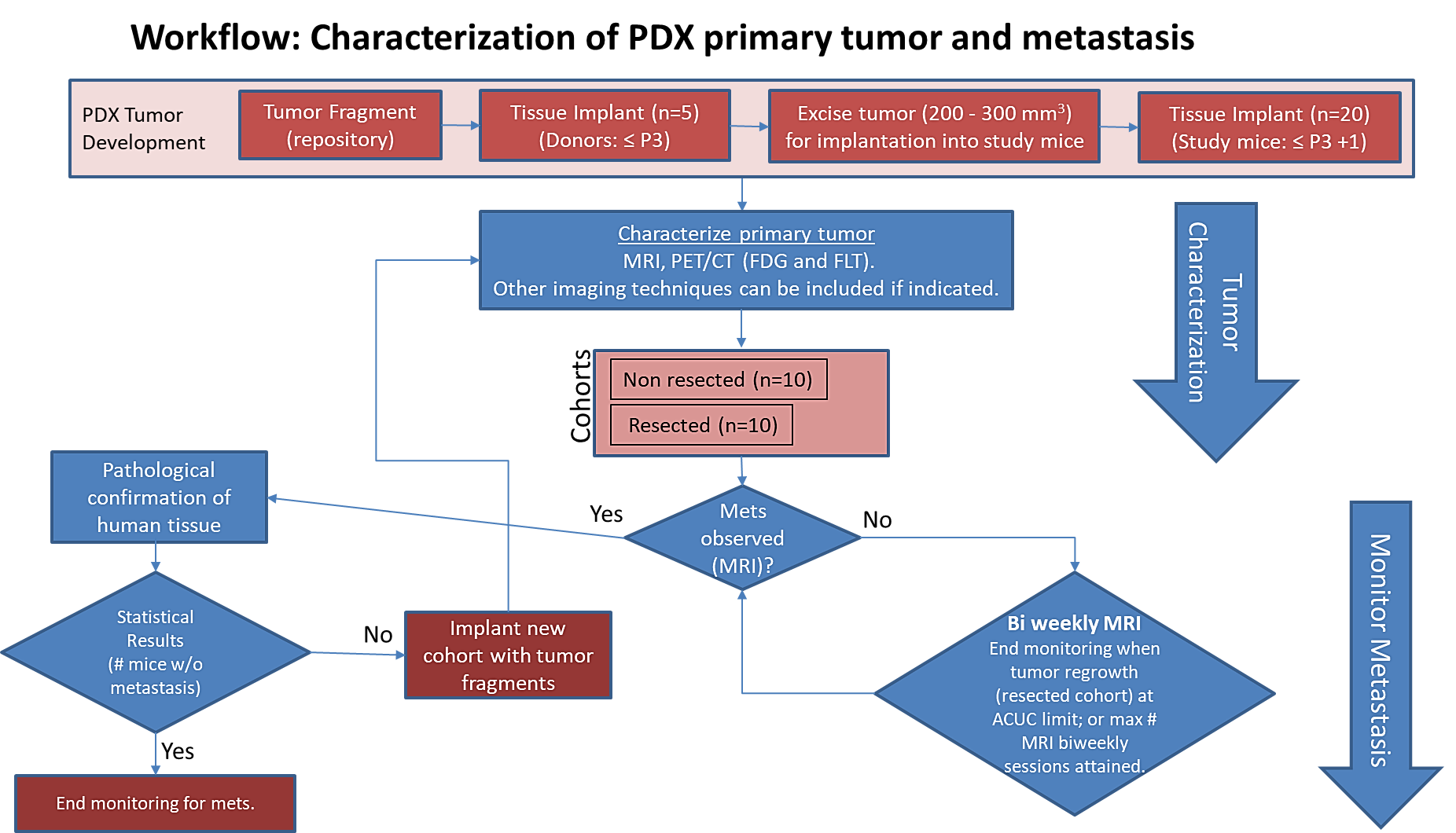
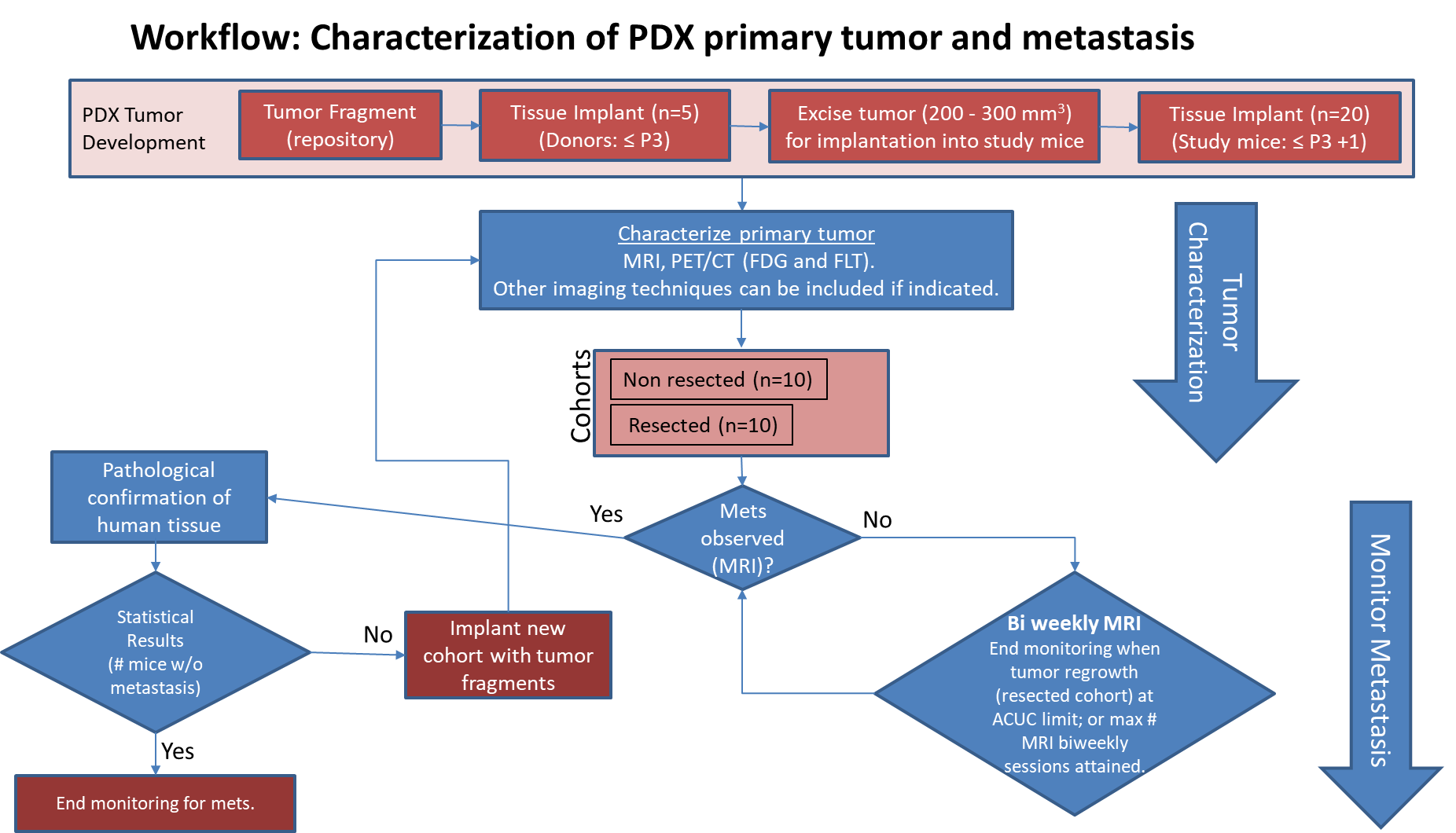
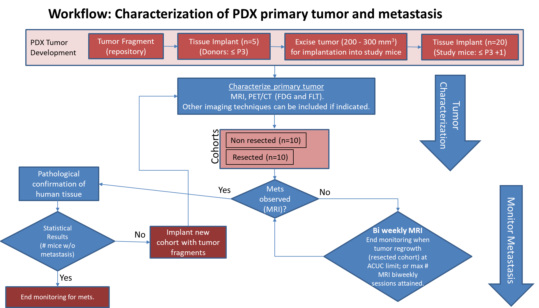
Pre-clinical animal models of spontaneous metastatic cancer are infrequent; the few that exist are resource intensive because determination of the presence of metastatic disease, metastatic burden, and response to therapy normally require multiple timed cohorts with animal sacrifice and extensive pathological examination. We identified and characterized a patient derived xenograft model with metastatic potential, melanoma...

Read More

PDMR-BL0293-F563

By
DOI: 10.7937/tcia.2019.b6u7wmqw | Image Collection

Pre-clinical animal models of spontaneous metastatic cancer are infrequent; the few that exist are resource intensive because determination of the presence of metastatic disease, metastatic burden, and response to therapy normally require animal sacrifice and extensive pathological examination. We recently identified and characterized a patient derived xenograft model with metastatic potential, bladder xenograft...

Read More

PDMR-521955-158-R4

By
DOI: 10.7937/q37d-vh79 | Image Collection

Pre-clinical animal models of spontaneous metastatic cancer are infrequent; the few that exist are resource intensive because determination of the presence of metastatic disease, metastatic burden, and response to therapy normally require multiple timed cohorts with animal sacrifice and extensive pathological examination. We identified and characterized a patient derived xenograft model with metastatic potential, adenocarcinoma...

Read More

PDMR-292921-168-R

By
DOI: 10.7937/TCIA.2020.PCAK-8Z10 | Image Collection

Pre-clinical animal models of spontaneous metastatic cancer are infrequent; the few that exist are resource intensive because determination of the presence of metastatic disease, metastatic burden, and response to therapy normally require multiple timed cohorts with animal sacrifice and extensive pathological examination. We identified and characterized a patient derived xenograft model with metastatic potential, adenocarcinoma...

Read More

PDMR-997537-175-T

By
DOI: 10.7937/TCIA.2020.BRY9-4N29 | Image Collection

Pre-clinical animal models of spontaneous metastatic cancer are infrequent; the few that exist are resource intensive because determination of the presence of metastatic disease, metastatic burden, and response to therapy normally require multiple timed cohorts with animal sacrifice and extensive pathological examination. We identified and characterized a patient derived xenograft model with metastatic potential, adenocarcinoma...

Read More