HNSCC

By
DOI: 10.7937/k9/tcia.2020.a8sh-7363 | Image Collection

This collection contains imaging, radiation therapy, and clinical data from 627 head and neck squamous cell carcinoma (HNSCC) patients at MD Anderson Cancer Center.  Researchers at MDACC analyzed these patients' data as part of two separate research projects. 

Read More

CODEX-IMAGING-OF-HCC

By
DOI: 10.7937/BH0R-Y074 | Image Collection

Introduction:

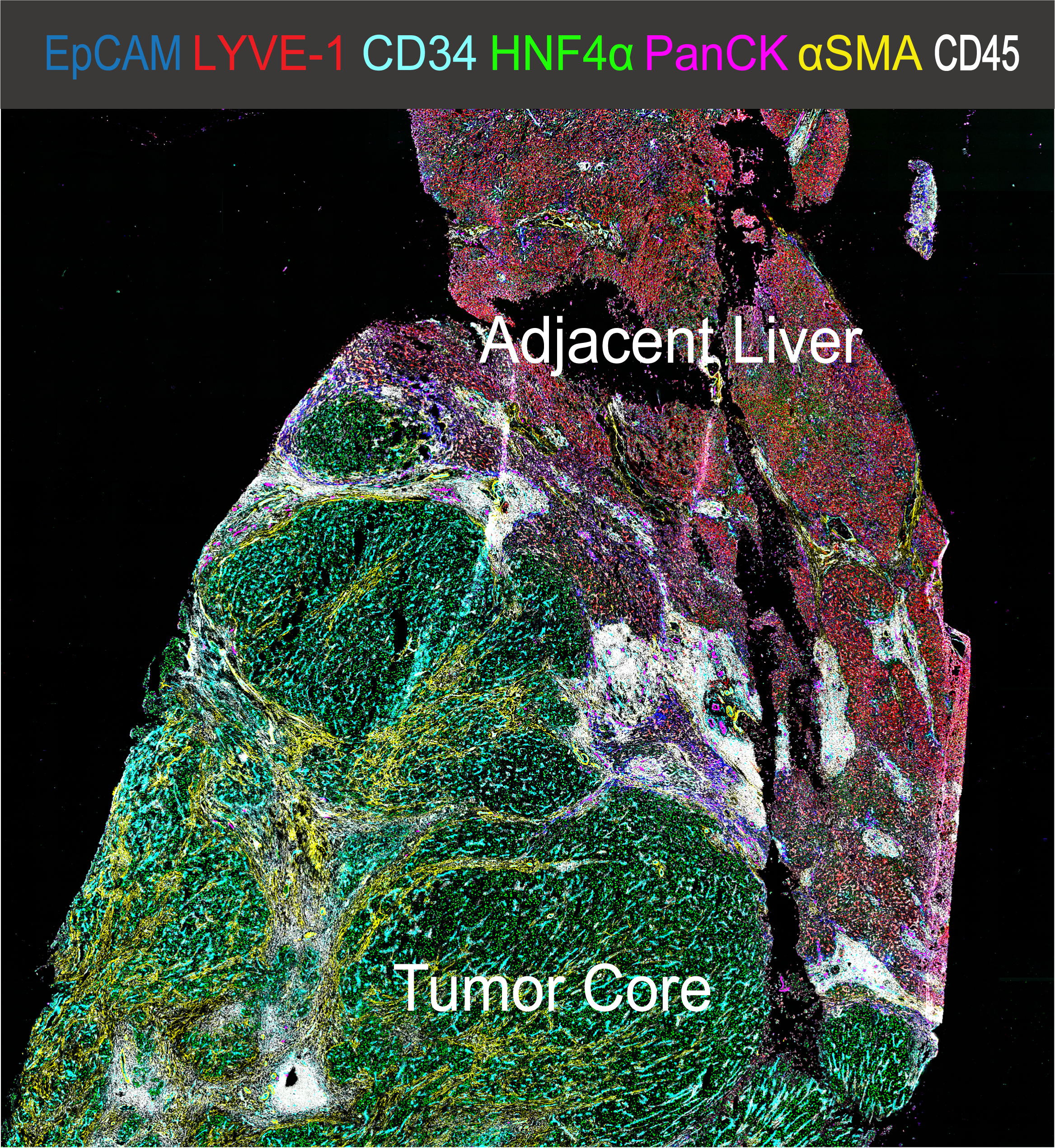
Hepatocellular Carcinoma (HCC) is a leading cause of cancer-related death and can be considered a prototype of inflammation-derived cancer arising from chronic liver injury. The cell composition of the HCC tumor immune microenvironment (TiME) has a major impact on cancer biology as the TiME can have divergent capacities on tumor initiation, progress, and response to therapy. Recent development...

Read More

CRC_FFPE-CODEX_CELLNEIGHS

By
DOI: 10.7937/TCIA.2020.FQN0-0326 | Image Collection

We have used CODEX to image 56 proteins simultaneously in 140 tissue regions from the tumor invasive front of 35 advanced-stage colorectal cancer (CRC) patients (17 patients with Crohn's-like reaction (CLR) - leading to high amount of tertiary lymphoid structures (TLS); and 18 patients with diffuse inflammatory infiltration (DII) and no TLS). These patients were selected from an initial cohort of 715 CRC patients....

Read More

HER2-TUMOR-ROIS

By
DOI: 10.7937/E65C-AM96 | Image Collection

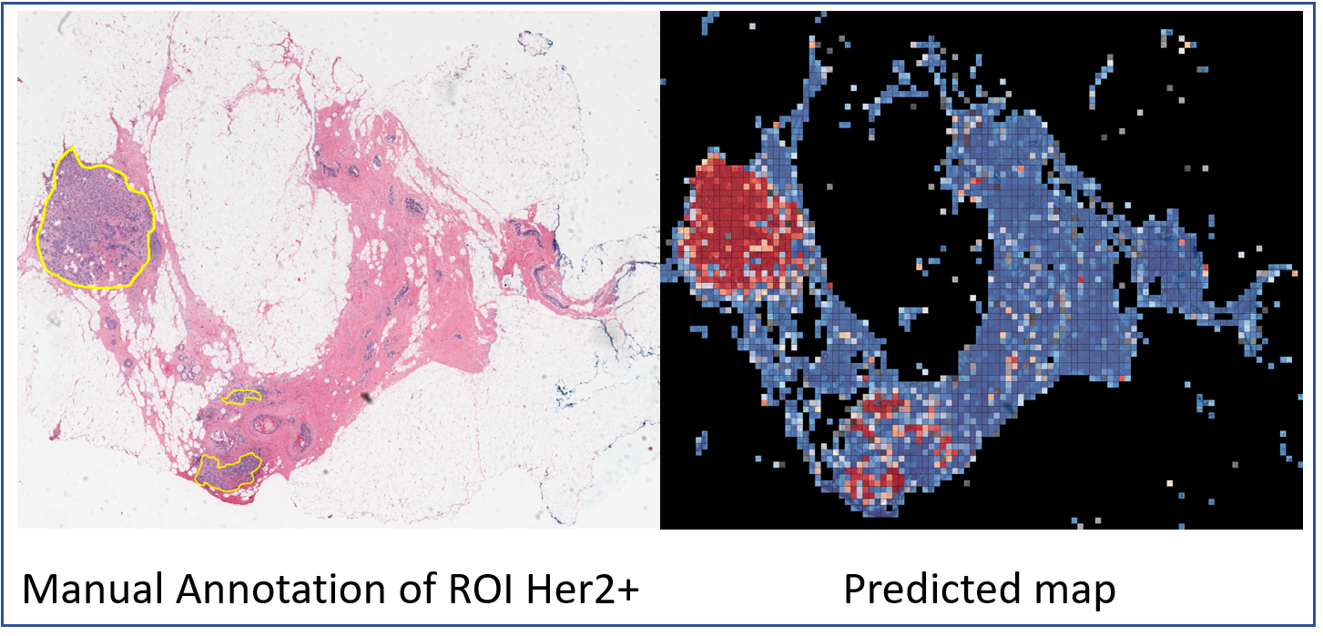
The current standard of care for many patients with HER2-positive breast cancer is neoadjuvant chemotherapy in combination with anti-HER2 agents, based on HER2 amplification as detected by in situ hybridization (ISH) or protein immunohistochemistry (IHC). However, hematoxylin & eosin (H&E) tumor stains are more commonly available, and accurate prediction of HER2 status and anti-HER2 treatment response from...

Read More

DLBCL-MORPHOLOGY

By
DOI: 10.7937/NVA3-N783 | Image Collection


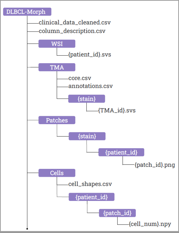
Diffuse Large B-Cell Lymphoma (DLBCL) is the most common non-Hodgkin lymphoma worldwide. DLBCL is fatal without treatment, but early detection and therapy can cure up to 70% of patients. The current best prognostic classification, the National Comprehensive Cancer Network International Prognostic Index, is insufficient to guide therapeutic decision-making for individual patients. No tumor-intrinsic prognostication...

Read More

HEAD-NECK-RADIOMICS-HN1

By
DOI: 10.7937/tcia.2019.8kap372n | Image Collection

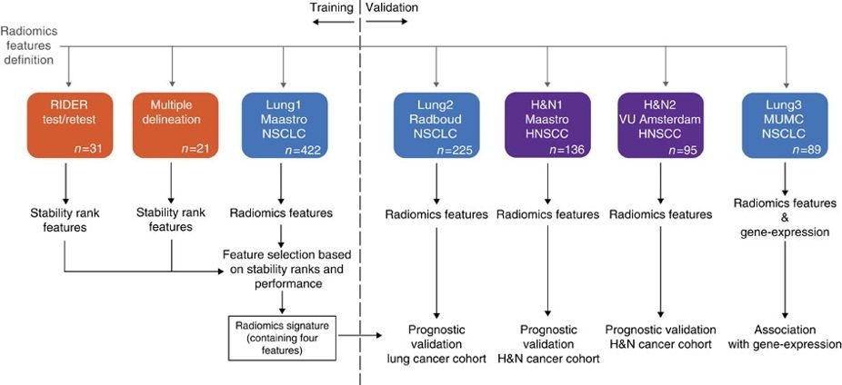
This collection contains clinical data and computed tomography (CT) from 137 head and neck squamous cell carcinoma (HNSCC) patients treated by radiotherapy. For these patients a pre-treatment CT scan was manual delineated by an experienced radiation oncologist of the 3D volume of the gross tumor volume. This dataset refers to the "H&N1" dataset of the study published in Nature Communications (http://doi.org/10.1038/ncomms5006). At...

Read More

HEAD-NECK-PET-CT

By
DOI: 10.7937/K9/TCIA.2017.8oje5q00 | Image Collection

This collection contains FDG-PET/CT and radiotherapy planning CT imaging data of 298  patients from four different institutions in Québec with histologically proven head-and-neck cancer (H&N) All patients had pre-treatment FDG-PET/CT scans between April 2006 and November 2014, and within a median of 18 days (range: 6-66) before treatment. Dates in the TCIA images have been changed in the interest of de-identification;...

Read More

HEAD-NECK-CETUXIMAB

By
DOI: 10.7937/K9/TCIA.2015.7AKGJUPZ | Image Collection

This collection combines advanced molecular imaging treatment response assessment through pre- and post-treatment FDG PET/CT scans with therapy of advanced head and neck cancer, including chemo-radiation therapy with and without addition of an EGFR inhibitor molecular targeted agent (Cetuximab). 

The Head-Neck Cetuximab collection consists of a subset of image data from RTOG 0522, which was randomized phase...

Read More

HNSCC-3DCT-RT

By
DOI: 10.7937/K9/TCIA.2018.13upr2xf | Image Collection

Researchers will have the unique opportunity to visualize how head-and-neck squamous cell carcinoma (HNSCC) patients’ 3D CT image data changes with radiotherapy between pre-treatment, mid-treatment, and post-treatment. They will also have the advantage to receive additional information such as a) photon energy, b) number of arcs for volumetric modulated arc radiotherapy (VMAT) therapy, c) tumor volume and location,...

Read More

GLIS-RT

By
DOI: 10.7937/TCIA.T905-ZQ20 | Image Collection

This collection consists of 230 cases of glioblastoma and low-grade glioma patients treated with surgery and adjuvant radiotherapy at Massachusetts General Hospital. The patients underwent routine post-surgical MRI examination by acquiring two MR sequences, contrast enhanced 3D-T1 and 2D multislice-T2 FLAIR required to define target volumes for radiotherapy treatment. CT scans were acquired after diagnostic imaging...

Read More