
GLIS-RT

By
DOI: 10.7937/TCIA.T905-ZQ20 | Image Collection

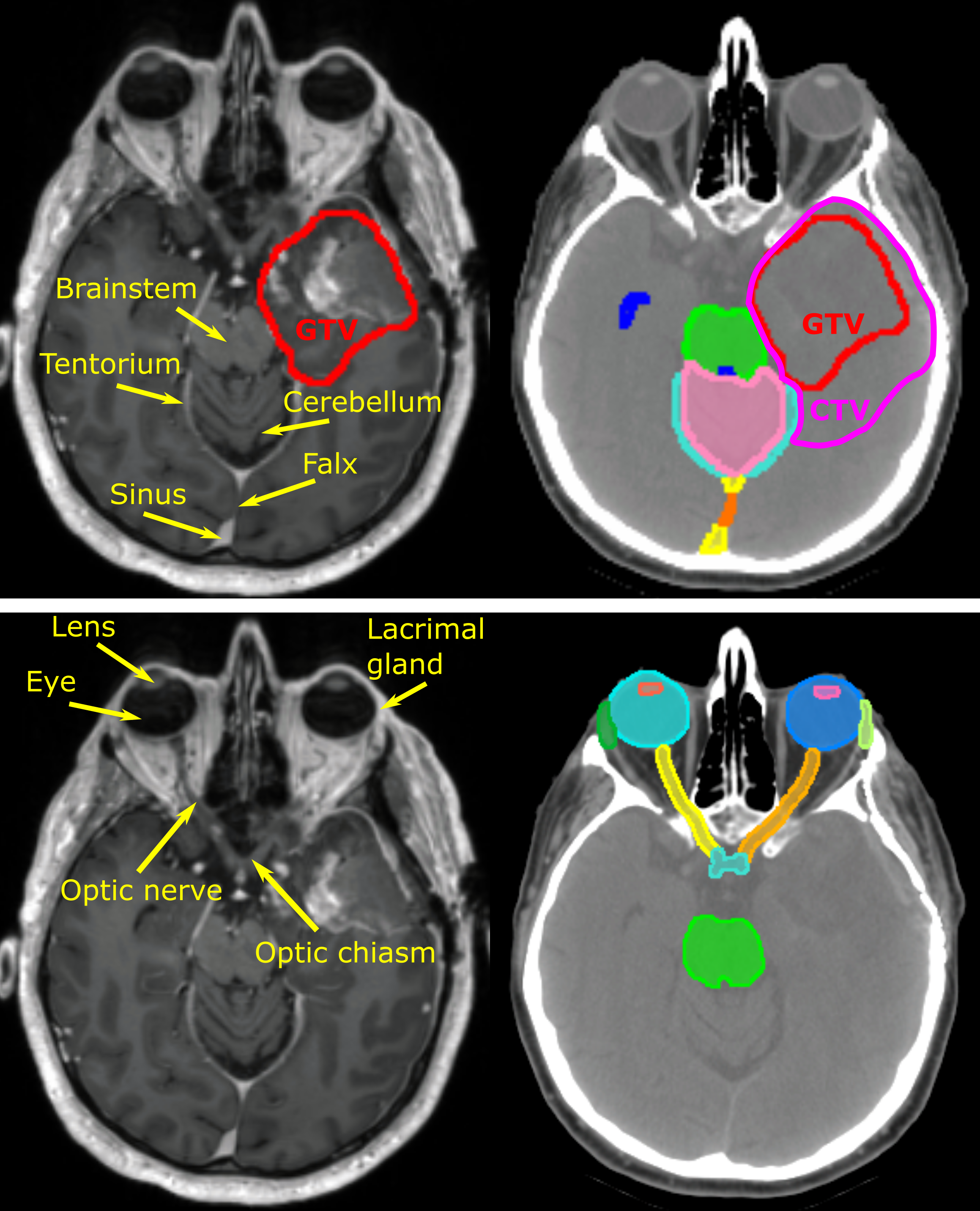
This collection consists of 230 cases of glioblastoma and low-grade glioma patients treated with surgery and adjuvant radiotherapy at Massachusetts General Hospital. The patients underwent routine post-surgical MRI examination by acquiring two MR sequences, contrast enhanced 3D-T1 and 2D multislice-T2 FLAIR required to define target volumes for radiotherapy treatment. CT scans were acquired after diagnostic imaging...

Read More

QIN-BRAIN-DSC-MRI

By
DOI: 10.7937/K9/TCIA.2016.5DI84Js8 | Image Collection

This collection contains MR images of both low and high grade glial brain lesions. Data includes post-contrast T1w images with co-registered volumes of dynamic susceptibility contrast (DSC) MR images in DICOM format. Binary regions of interest are also included, in DICOM format, of the lesion, arterial input function, normal appearing white matter, normal appearing cerebral cortex, and whole brain. The data was provided...

Read More

GBM-DSC-MRI-DRO

By
DOI: 10.7937/TCIA.2020.RMWVZWIX | Image Collection

The standardization of dynamic susceptibility contrast (DSC)-magnetic resonance imaging (MRI) has been confounded by a lack of consensus on DSC-MRI methodology for preventing potential relative cerebral blood volume (CBV) inaccuracies, including the choice of acquisition protocols and postprocessing algorithms. Therefore, a digital reference object (DRO) was developed using physiological and kinetic parameters...

Read More

GAMMAKNIFE-HIPPOCAMPAL

By
DOI: 10.7937/Q967-X166 | Image Collection

This collection is comprised of 390 patients who presented with vestibular schwannoma (VS, n=73), trigeminal neuralgia (TGN, n=119) or metastatic disease (M, n=198, 4-26 metastases) and were subsequently treated with Gamma Knife (Elekta AB, Stockholm, Sweden) stereotactic radiosurgery. For each patient, the treatment indication is designated with a suffix on their patient ID (VS, TGN or M).

All patients are provided...

Read More

PROSTATE-FUSED-MRI-PATHOLOGY

By
DOI: 10.7937/k9/TCIA.2016.tlpmr1am | Image Collection

This collection comprises a total of 28 3 Tesla T1-weighted, T2-weighted, Diffusion weighted and Dynamic Contrast Enhanced prostate MRI along with accompanying digitized histopathology (H&E stained) images of corresponding radical prostatectomy specimens. The MRI scans also have a mapping of extent of prostate cancer on them [10.1002/jmri.24975]. Each surgically...

Read More

LUNG-FUSED-CT-PATHOLOGY

By
DOI: 10.7937/k9/tcia.2018.smt36lpn | Image Collection

This is the first attempt of mapping the extent of Invasive Adenocarcinoma onto in vivo lung CT. The mappings constitute ground truth of disease and may be used to further investigate the imaging signatures of Invasive Adenocarcinoma in ground glass pulmonary nodules. Patient with small ground glass nodules with >2 histology slices per nodule were included. Patients with solid large nodules (>40mm), with <3...

Read More

EXACT

By
DOI: 10.7937/2023.na2x-j031 | Image Collection

Accurate pre-therapeutic imaging of the tumour and regional lymph nodes, is - in addition to clinical examination and assessment for distant metastases -essential for prognostication and guiding treatment. Nodal staging has been validated as an important independent prognostic factor [EORTC22861; RTOG9811] and the nodal stage as well as the precise location of involved lymph nodes (mesorectum, presacral space, internal...

Read More

DUKE-BREAST-CANCER-MRI

By
DOI: 10.7937/TCIA.e3sv-re93 | Image Collection

Breast MRI is a common image modality to assess the extent of disease in breast cancer patients. Recent studies show that MRI has a potential in prognosis of patients’ short and long-term outcomes as well as predicting pathological and genomic features of the tumors. However, large, well annotated datasets are needed to make further progress in the field. We share such a dataset here.

In terms of design, the...

Read More

HUNGARIAN-COLORECTAL-SCREENING

By
DOI: 10.7937/TCIA.9CJF-0127 | Image Collection

In this study, 200 digital whole-slide images are published which were collected via hematoxylin-eosin stained colorectal biopsy. This dataset contains the raw MIRAX (mrxs) formatted data. The samples were selected from the archives of the 2nd Department of Pathology of Semmelweis University, Budapest and were scanned with a 3DHistech Pannoramic 1000 Digital Slide Scanner at the highest available, 40x magnification....

Read More

C4KC-KITS

By
DOI: 10.7937/TCIA.2019.IX49E8NX | Image Collection

This collection contains CT scans and segmentations from subjects from the training set of the 2019 Kidney and Kidney Tumor Segmentation Challenge (KiTS19). The challenge aimed to accelerate progress in automatic 3D semantic segmentation by releasing a dataset of CT scans for 210 patients with manual semantic segmentations of the kidneys and tumors in the corticomedullary...

Read More