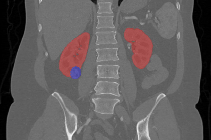
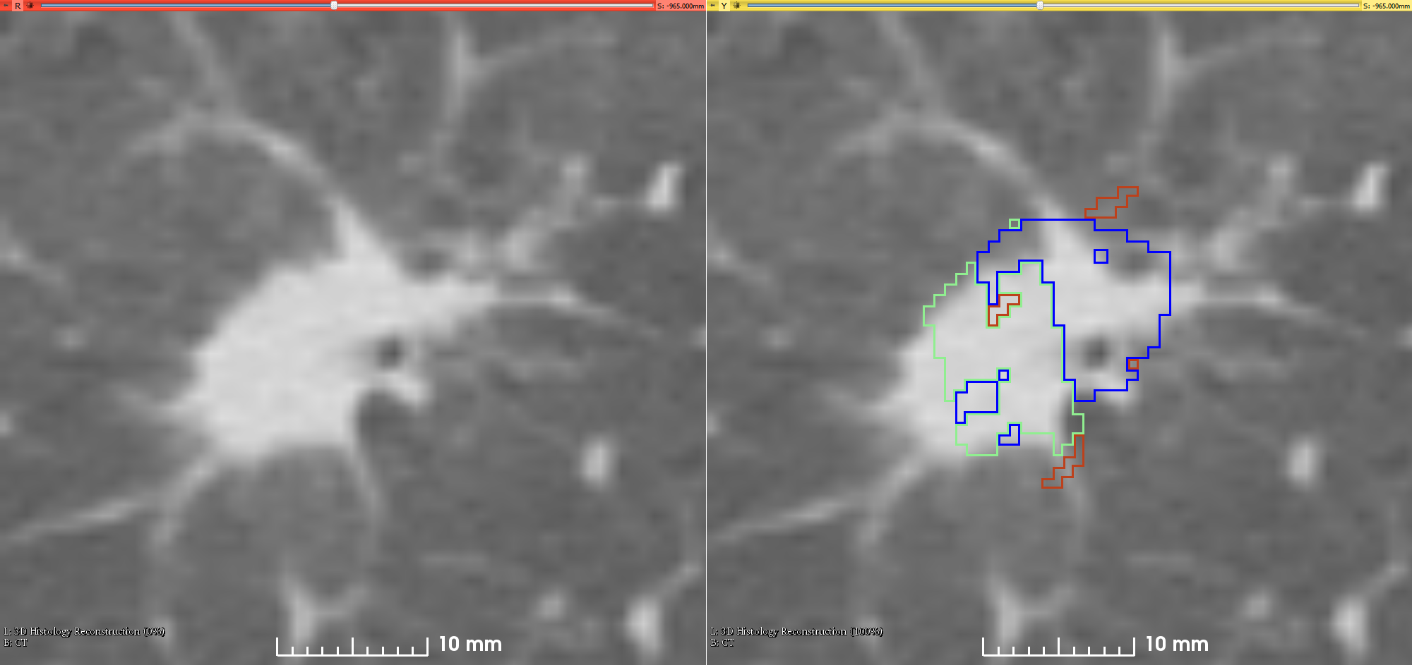
The Lung Image Database Consortium image collection (LIDC-IDRI) consists of diagnostic and lung cancer screening thoracic computed tomography (CT) scans with marked-up annotated lesions. It is a web-accessible international resource for development, training, and evaluation of computer-assisted diagnostic (CAD) methods for lung cancer detection and diagnosis. Initiated by the National Cancer Institute (NCI), further...
This data collection consists of images acquired during chemoradiotherapy of 20 locally-advanced, non-small cell lung cancer patients. The images include four-dimensional (4D) fan beam (4D-FBCT) and 4D cone beam CT (4D-CBCT). All patients underwent concurrent radiochemotherapy to a total dose of 64.8-70 Gy using daily 1.8 or 2 Gy fractions.
4D-FBCT images were acquired on a 16-slice helical CT scanner (Brilliance...
This CBIS-DDSM (Curated Breast Imaging Subset of DDSM) is an updated and standardized version of the Digital Database for Screening Mammography (DDSM). The DDSM is a database of 2,620 scanned film mammography studies. It contains normal, benign, and malignant cases with verified pathology information. The scale of the database...
This dataset consists of 140 computed tomography (CT) scans, each with five organs labeled in 3D: lung, bones, liver, kidneys and bladder. The brain is also labeled on the minority of scans which show it.
Patients were included based on the presence of lesions in one or more of the labeled organs. Most of the images exhibit liver lesions, both benign and malignant. Some also exhibit metastatic disease in other...

For this study, 20 lung cancer patients underwent exhale/inhale breath hold CT (BHCT), free-breathing four-dimensional CT (4DCT) and Galligas PET ventilation scans in a single session on a combined 4DPET/CT scanner. The purpose of the study was to enable comparisons between: (i) CT ventilation images derived from exhale/inhale BHCT scans, (ii) CT ventilation images derived from free-breathing 4DCT scans, and (iii)...

There currently is a dearth of phantom scans on large samples. This data collection contains one physical phantom, imaged across three protocols, on 100 scanners. This provides population data that can be used to quantify inter-scanner variability. This data can be used to determine how robust specific radiomics or other quantitative imaging signatures are.
Protocol
Computed tomography scans were acquired...

These retrospective NIfTI image datasets consists of unenhanced chest CTs:
- First dataset - from 632 patients with COVID-19 infections at initial point of care, and
- Second dataset - a second set of 121 CTs from 29 patients with COVID-19 infections with serial / sequential CTs.
The initial images for both datasets were acquired at the point of care in an outbreak setting from patients...
The National CT Colonography Trial (ACRIN 6664) collection contains 825 cases of CT colonography imaging with accompanying spreadsheets that provide polyp descriptions and their location within the colon segments. Additional information about the trial is available in the
This collection consists of 251 CT scans of Credence Cartridge Radiomic (CCR) phantom. This texture phantom was developed to investigate the feature robustness in the emerging field of radiomics. This phantom dataset was acquired on 4-8 CT scanners using a set of imaging parameters (e.g., reconstruction Field of View, Slice thickness, reconstruction kernels, mAs, and Pitch). A controlled scanning approach was employed...

This collection consists of 17 CT scans of the Credence Cartridge Radiomics (CCR) phantom, which was designed for use in studies of texture feature robustness. The scans were acquired at four medical centers using each center’s chest protocol and were taken using GE (7 scans), Philips (5 scans), Siemens (2 scans), and Toshiba (3 scans) scanners. The CCR phantom has 10 cartridges, each with a unique texture, Fig 1....