HNC-IMRT-70-33

By
DOI: 10.7937/ahqh-xc79 | Image Collection

This collection includes data from 211 patients who presented with head and neck cancer and were treated using radiation therapy at a single institution using 6 MV Intensity Modulated Radiation Therapy (IMRT) from a linear accelerator. All patients were prescribed identical prescriptions of 70 Gray (Gy) in 33 fractions for the primary planning target volume (PTV) and one, none, or a combination of integrated boost...

Read More

CPTAC-HNSCC

By
DOI: 10.7937/K9/TCIA.2018.UW45NH81 | Image Collection

This collection contains subjects from the National Cancer Institute’s Clinical Proteomic Tumor Analysis Consortium Head-and-Neck cancer (CPTAC-HNSCC) cohort. CPTAC is a national effort to accelerate the understanding of the molecular basis of cancer through the application of large-scale proteome and genome analysis, or proteogenomics. Radiology...

Read More

RADCURE

By
DOI: 10.7937/J47W-NM11 | Image Collection

The RADCURE dataset was collected clinically for radiation therapy treatment planning and retrospectively reconstructed for quantitative imaging research.  

Inclusion: The dataset used for this study consists of 3,346 head and neck cancer CT image volumes collected from 2005-2017 treated with definitive RT at the University Health Network (UHN) in Toronto, Canada

Acquisition and...

Read More

PRETREAT-METSTOBRAIN-MASKS

By
DOI: 10.7937/6be1-r748 | Image Collection

Cohort Selection:

Patient images were collected from three sources: the Yale New Haven Health database (2013-2021), the Yale tumor board registry (2021), and the Yale gamma knife registry (2017-2021).

Inclusion/exclusion criteria:

Inclusion criteria included a pathologically proven diagnosis of brain metastasis and availability of a pretreatment scan with standard MRI sequences...

Read More

BREAST-LESIONS-USG

By
DOI: 10.7937/9WKK-Q141 | Image Collection

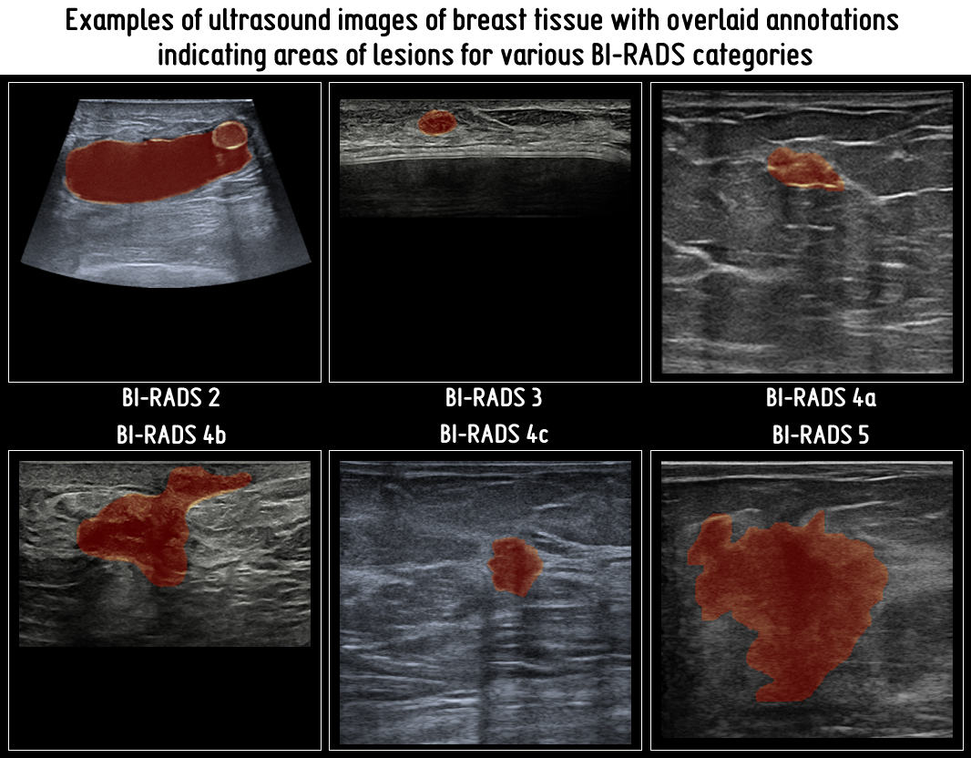
This dataset consists of 256 breast ultrasound scans collected from 256 patients and 266 benign and malignant segmented lesions.  It includes patient-level labels, image-level annotations, and tumor-level labels with all cases confirmed by follow-up care or biopsy result. Each scan was manually annotated and labeled by a radiologist experienced in breast ultrasound examination. In particular, each tumor was identified...

Read More

HE-VS-MPM

By
DOI: 10.7937/3fyc-ac78 | Image Collection

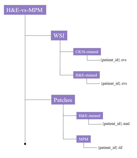
Ductal carcinoma in situ with microinvasion (DCISM) is a challenging subtype of breast cancer with controversial invasiveness and prognosis. Accurate diagnosis of DCISM from ductal carcinoma in situ (DCIS) is crucial for optimal treatment and improved clinical outcomes. This dataset provides histopathology images and paired CK5/6 immunohistochemical staining images from patients with DCISM, as well as multiphoton microscopy...

Read More

ADVANCED-MRI-BREAST-LESIONS

By
DOI: 10.7937/C7X1-YN57 | Image Collection

The application of deep-learning methods to breast MRI has a potential to improve diagnosis as well as predict pathological features of the tumors and their response to therapy, and is currently widely investigated. Standard contrast-enhanced MRI of the breast provides high sensitivity but variable specificity in detecting breast cancer, and may lead to excessive benign biopsies. Large, well annotated datasets are...

Read More

PROSTATE-ANATOMICAL-EDGE-CASES

By
DOI: 10.7937/QSTF-ST65 | Image Collection

In this single institution retrospective study, we reviewed 950 consecutive patients with prostate adenocarcinoma receiving definitive radiotherapy between 2011 and 2019, and identified among them 112 patients with anatomic variations (edge cases) seen on simulation CT and/or MRI imaging. These variations included hip arthroplasty, prostate median lobe hypertrophy, so-called “droopy” seminal vesicles, presence...

Read More

QIN-BREAST

By
DOI: 10.7937/K9/TCIA.2016.21JUEBH0 | Image Collection

This collection contains longitudinal PET/CT and quantitative MR images collected for the purpose of studying treatment assessment in breast cancer in the neoadjuvant setting. Images were acquired at three time points: prior to the start of treatment (t1), after the first cycle of treatment (t2), and either after the second cycle of treatment or at the completion of all treatments (prior to surgery) (t3). The PET/CT...

Read More