Read More
Microscopic images were captured from bone marrow aspirate slides of patients diagnosed with B-lineage Acute Lymphoid Leukemia (B-ALL) and Multiple Myeloma (MM) as per the standard guidelines. Slides were stained using Jenner-Giemsa stain. Images were captured at 1000x magnification using Nikon Eclipse-200 microscope equipped with a digital camera. Images were captured in raw BMP format with a size of 2560x1920 pixels....
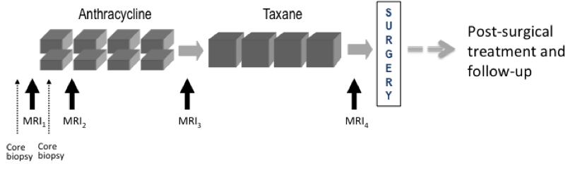
This collection contains longitudinal DCE MRI studies of 64 patients undergoing neoadjuvant chemotherapy (NACT) for invasive breast cancer.
Patient population
This pilot study to investigate the use of serial DCE MRI examinations during neoadjuvant chemotherapy for invasive breast cancer recruited 68 patients with stage II or III locally advanced breast cancer enrolled between...
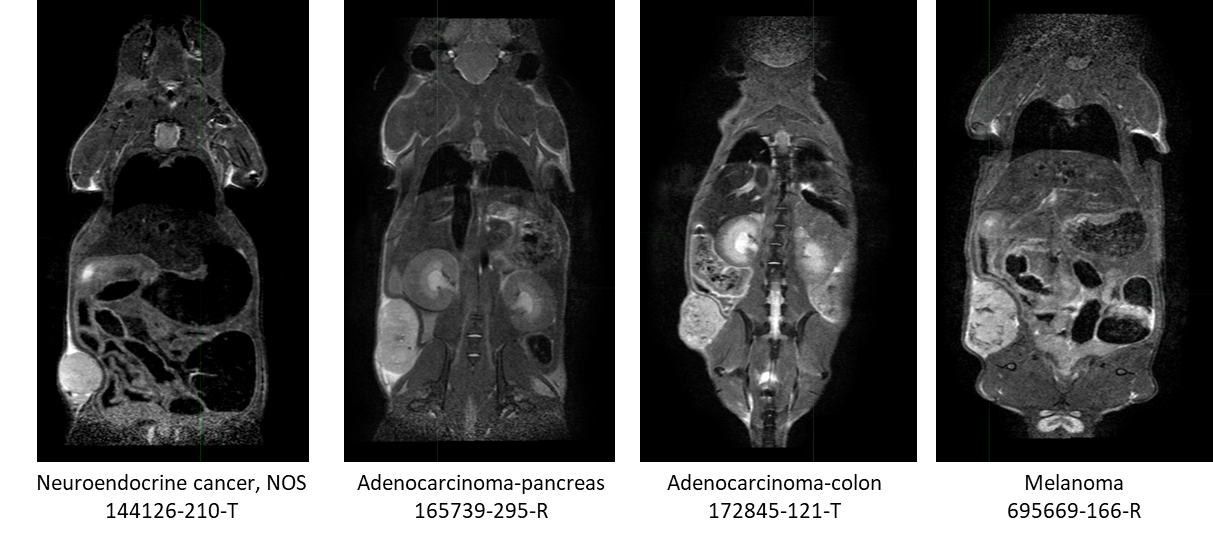
This collection contains serial non-contrast non-gated T2w MRI of 18 patient derived xenograft cancer models. 175 mice were imaged at multiple time points (514 total studies) for researchers to develop algorithms using neural networks, and classification techniques to improve tissue characterization (morphological changes) for the improvement in patient care through advances in precision medicine.
Characterization...
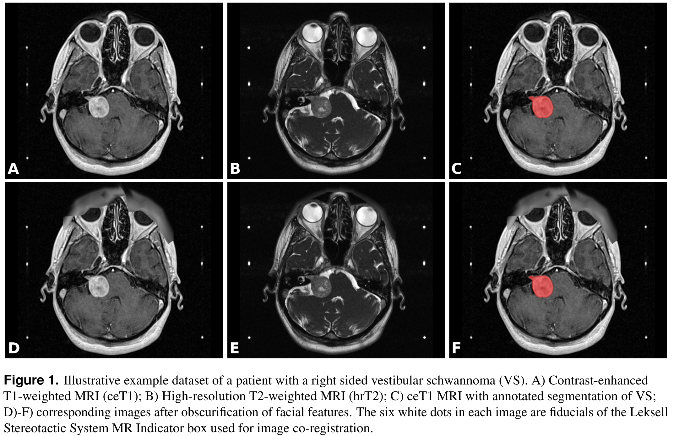
This collection contains a labeled dataset of MRI images collected on 242 consecutive patients with vestibular schwannoma (VS) undergoing Gamma Knife stereotactic radiosurgery (GK SRS). The structural images included contrast-enhanced T1-weighted (ceT1) images and high-resolution T2-weighted (hrT2) images. Each imaging dataset is accompanied by the patient’s radiation therapy (RT) dataset including the RTDose, RTStructures...
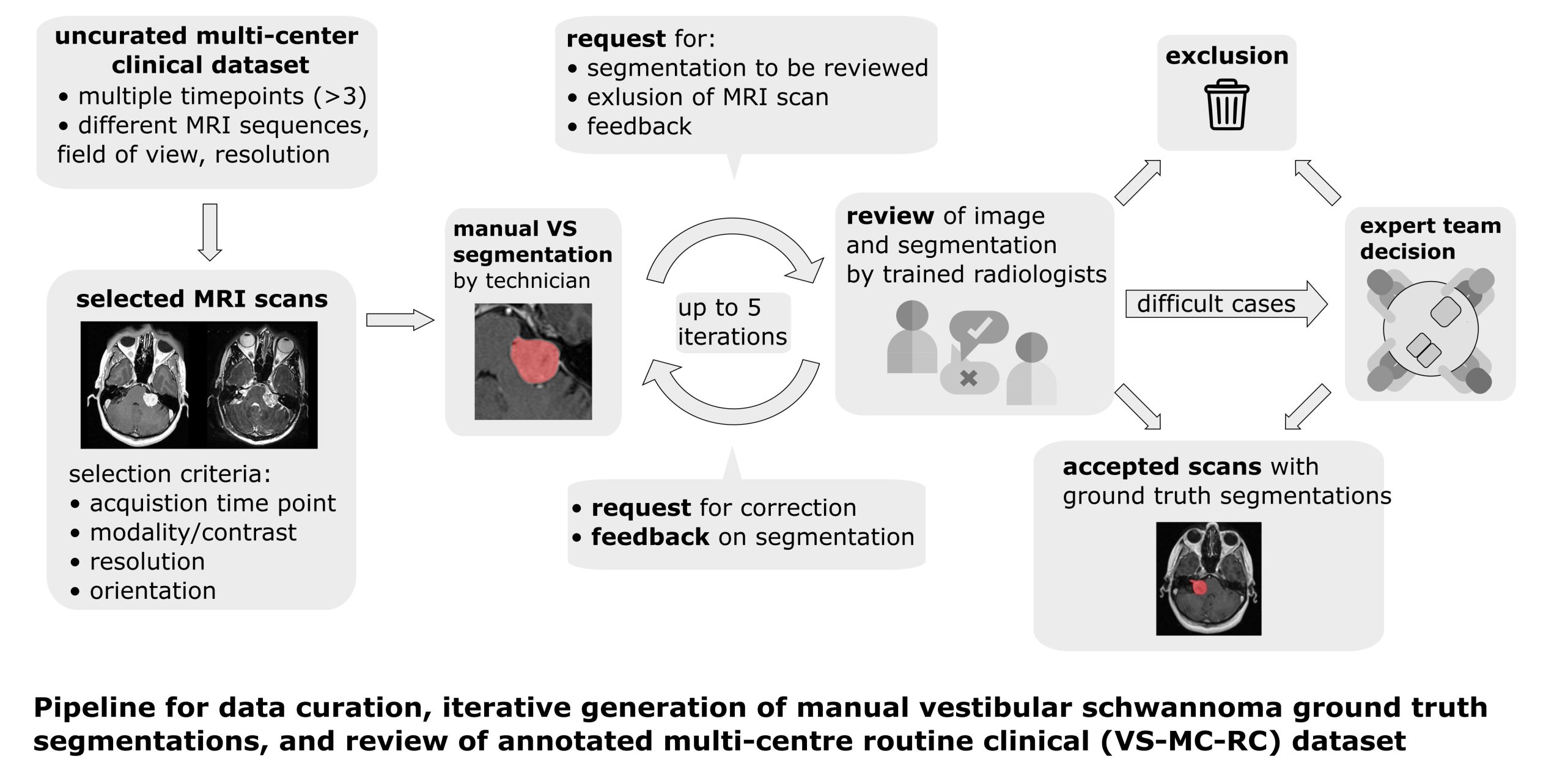
The Vestibular-Schwannoma-MC-RC (VS-MC-RC) dataset contains longitudinal MRI scans of 160 patients with a single sporadic Vestibular Schwannoma (VS) from 10 medical sites in the United Kingdom. For all patients either a T1-weighted (T1w) or T2-weighted (T2w) scan, or both, are available. The dataset provides binary segmentations of the VS for all scan sessions. The dataset comprises up to three time points for each...
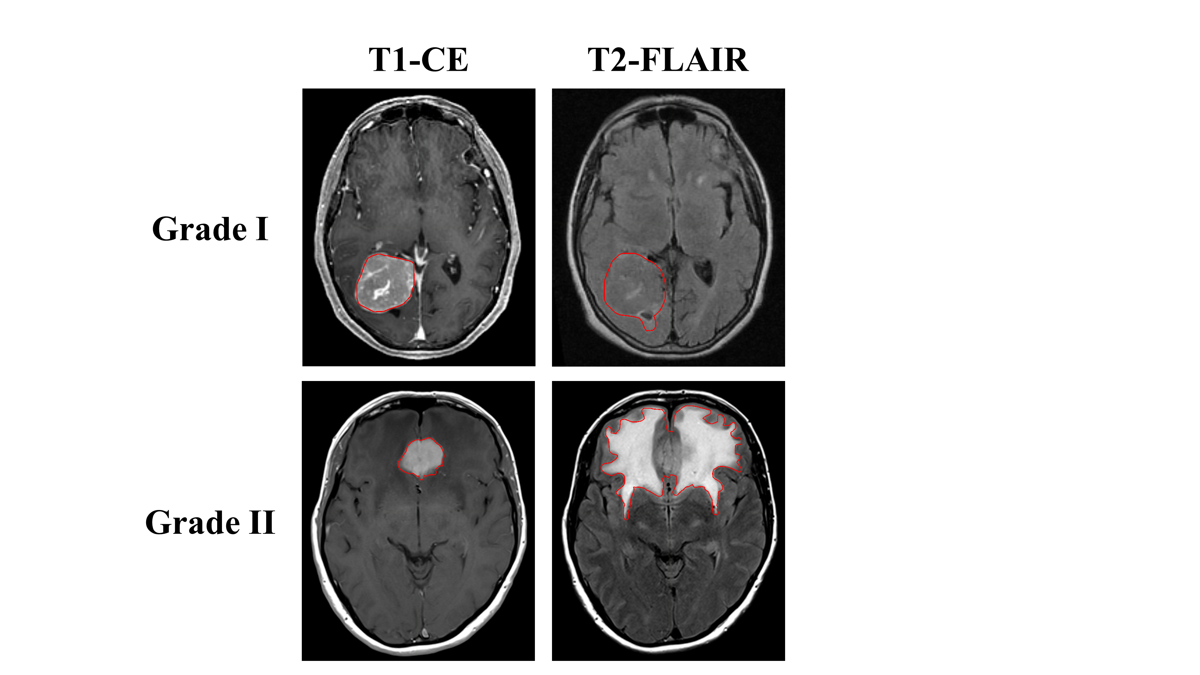
The study included 96 consecutive treatment naïve patients with intracranial meningiomas treated with surgical resection from 2010 to 2019. All patients had pre-operative T1, T1-CE, and T2-FLAIR MR images with subsequent subtotal or gross total resection of pathologically confirmed grade I or grade II meningiomas. A neuropathology team reviewed histopathology, including two subspecialty trained neuropathologists and...