BREAST-MRI-NACT-PILOT

DOI: 10.7937/K9/TCIA.2016.QHsyhJKy | Image Collection

This collection contains longitudinal DCE MRI studies of 64 patients undergoing neoadjuvant chemotherapy (NACT) for invasive breast cancer. 

Patient population

This pilot study to investigate the use of serial DCE MRI examinations during neoadjuvant chemotherapy for invasive breast cancer recruited 68 patients with stage II or III locally advanced breast cancer enrolled between...

[…] undergoing neoadjuvant chemotherapy (NACT) for invasive breast cancer.  Patient population […]

CROWDS-CURE-2018

DOI: 10.7937/TCIA.2019.yk0gm1eb | Analysis Result

Description

The Cancer Imaging Archive (TCIA) has procured substantial troves of image data, which could serve as valuable training sets for improving machine learning algorithms. However, these datasets lack consistent lesion annotations. To address this issue, the Cancer Imaging Informatics Lab at the...

[…] address this issue, the Cancer Imaging Informatics Lab at the Frederick National […]

CDD-CESM

DOI: 10.7937/29kw-ae92 | Image Collection

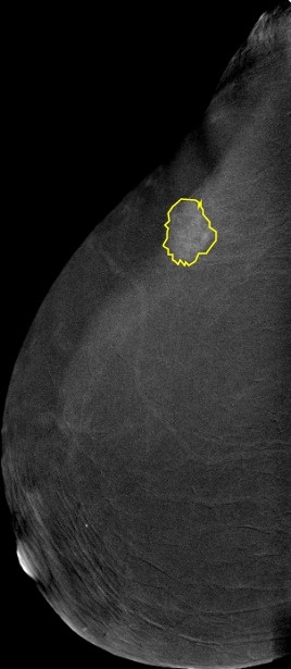
Deep learning (DL) has a promising potential in reducing the workload of radiologists and helping them provide a more accurate diagnosis. However, fully annotated and large-sized datasets are required. This dataset is a collection of 2,006 high-resolution Contrast-enhanced spectral mammography (CESM) images with annotations and medical reports. 

Acquisition protocol: 

CESM is done using...

[…] (DL) has a promising potential in reducing the workload of radiologists and helping them provide a more accurate diagnosis. However, fully annotated and large-sized datasets are required. This dataset is a collection of 2,006 high-resolution Contrast-enhanced spectral mammography (CESM) images with annotations and medical reports.  Acquisition protocol:  CESM is done using the standard digital mammography equipment, with additional software that performs dual-energy image acquisition. Two minutes after intravenously injecting the patient with non-ionic low-osmolar iodinated contrast material (dose: 1.5 mL/kg), craniocaudal (CC) and mediolateral oblique (MLO) views are obtained. Each view comprises two exposures, one with low energy (peak kilo-voltage values ranging from 26 to 31kVp) and one with high energy (45 to 49 kVp). Low and high-energy images are then recombined and subtracted through appropriate image processing to suppress the background breast parenchyma. A complete examination is carried out in about 5-6 minutes. Image preprocessing: The images were converted from DICOM to JPEG using RadiAnt with best 100% image quality (lossless).  They have an average of 2355 x 1315 pixels. Supporting data: Full medical reports are also provided for each case (DOCX) along with manual segmentation annotation for the abnormal findings in each image (CSV file).  Each image with its corresponding manual annotation (breast composition, mass shape, mass margin, mass density, architectural distortion, asymmetries, calcification type, calcification distribution, mass enhancement pattern, non-mass enhancement pattern, non-mass enhancement distribution, and overall BIRADS assessment) is compiled into 1 Excel file.  https://www.robots.ox.ac.uk/~vgg/software/via/via.html was used for the segmentation annotation.  It can be used to show the annotations on the images by clicking on Annotation–> import annotations (from csv), and […]

ACRIN-NSCLC-FDG-PET

DOI: 10.7937/tcia.2019.30ilqfcl | Image Collection

Positron Emission Tomography Pre- and Post-treatment Assessment for Locally Advanced Non-small Cell Lung Carcinoma 

This was a multicenter clinical trial by the ACRIN Cooperative Group (now part of ECOG-ACRIN) and the RTOG Cooperative Group (now part of NRG) using FDG-PET imaging...

[…] for definitive concurrent chemoradiotherapy ( inoperable disease). Additional information about […]

HISTOLOGYHSI-GB

DOI: 10.7937/z1k6-vd17 | Image Collection

Hyperspectral imaging technology combines the main features of two existing technologies: conventional imaging and spectroscopy. Thus, hyperspectral cameras make it possible to analyze, at the same time and in a non-contact way, the morphological features and chemical composition of the objects captured. The information provided by hyperspectral imaging can be used to detect patterns, cells, or biomarkers to identify...

Summary Hyperspectral imag ing technology combines the main features of […]

CT-LYMPH-NODES

DOI: 10.7937/K9/TCIA.2015.AQIIDCNM | Image Collection

This collection consists of Computed Tomography (CT) images of the mediastinum and abdomen in which lymph node positions are marked by radiologists at the National Institutes of Health, Clinical Center. Radiologists at the Imaging Biomarkers and Computer-Aided Diagnosis Laboratory labeled a total...

[…] (CT) images of the mediast inum and abdomen in which […]

NLST

DOI: 10.7937/TCIA.HMQ8-J677 | Image Collection

Background: The aggressive and heterogeneous nature of lung cancer has thwarted efforts to reduce mortality from this cancer through the use of screening. The advent of low-dose helical computed tomography (CT) altered the landscape of lung-cancer screening, with studies indicating that low-dose CT detects many tumors at early stages. The National Lung Screening Trial (NLST) was conducted to determine...

[…] of lung-cancer screening, with studies indicating that low-dose CT detects […]

PANCREAS-CT

DOI: 10.7937/K9/TCIA.2016.tNB1kqBU | Image Collection

The National Institutes of Health Clinical Center performed 82 abdominal contrast enhanced 3D CT scans (~70 seconds after intravenous contrast injection in portal-venous) from 53 male and 27 female subjects.  Seventeen of the subjects are healthy kidney donors scanned prior to nephrectomy.  The remaining 65 patients were selected by a radiologist from patients who neither had major abdominal pathologies nor pancreatic...

Summary The National Institutes of Health Clinical Center performed 82 […]

ACRIN-FMISO-BRAIN

DOI: 10.7937/K9/TCIA.2018.vohlekok | Image Collection

The objective of the ACRIN 6684 multi-center clinical trial was to determine the association of baseline FMISO PET uptake (maximal tumor to blood ratio, hypoxic volume) and MRI parameters (Ktrans, CBV) with overall survival, time to disease progression, and 6-month progression free survival in participants with newly diagnosed glioblastoma multiforme (GBM). The trial also collected standard brain...

[…] right, and can be found online ( Protocol-ACRIN 6684 Amendment […]