ACRIN-FMISO-BRAIN
online ( Protocol-ACRIN 6684 Amendment […]

RSNA-ASNR-MICCAI-BRATS-2021
This dataset includes brain MRI scans of adult brain glioma patients, comprising of 4 structural modalities (i.e., T1, T1c, T2, T2-FLAIR) and associated manually generated ground truth labels for each tumor sub-region (enhancement, necrosis, edema), as well as their MGMT promoter methylation status. These scans are a collection of data from existing TCIA collections, but also cases provided by individual institutions...
This dataset includes brain MRI scans of adult brain glioma […]
RADIOMICS-TUMOR-PHENOTYPES
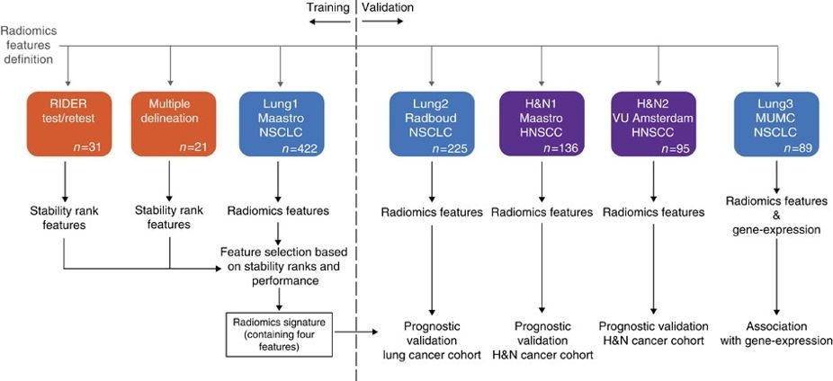
This data applies a radiomic approach to computed tomography data of 1,019 patients with lung or head-and-neck cancer which are described in Nature Communications (http://doi.org/10.1038/ncomms5006). The various arms of the study are represented in TCIA as distinct Collections including NSCLC-Radiomics (Lung1), […] head-and-neck cancer which are described in Nature Communications (http://doi.org/10.1038/ncomms5006). The […]
LUMINALB-BREAST-MR-ENHANCEMENT
Purpose
To investigate associations between breast cancer molecular subtype and semiautomatically extracted magnetic resonance (MR) imaging features.Materials and Methods
Imaging and genomic data from the Cancer Genome Atlas and the Cancer Imaging Archive for 48 patients with breast cancer from four institutions in the United States were used in this institutional review board approval-exempt study....[…] background parenchyma. (c) RSNA, 2014 Online supplemental material is available […]
Disclaimer Page
[…] system is detailed in the online help documentation accessible from […]
CT-ORG
This dataset consists of 140 computed tomography (CT) scans, each with five organs labeled in 3D: lung, bones, liver, kidneys and bladder. The brain is also labeled on the minority of scans which show it.
Patients were included based on the presence of lesions in one or more of the labeled organs. Most of the images exhibit liver lesions, both benign and malignant. Some also exhibit metastatic disease in other...
[…] each with five organs labeled in 3D: lung, bones, liver, […]
NSCLC-RADIOMICS-GENOMICS
This collection contains images from 89 non-small cell lung cancer (NSCLC) patients that were treated with surgery. For these patients pretreatment CT scans, gene expression, and clinical data are available. This dataset refers to the Lung3 dataset of the study published in Nature Communications.
In short, this publication...
Summary This collection conta ins images from 89 non-small cell […]
CBIS-DDSM
This CBIS-DDSM (Curated Breast Imaging Subset of DDSM) is an updated and standardized version of the Digital Database for Screening Mammography (DDSM). The DDSM is a database of 2,620 scanned film mammography studies. It contains normal, benign, and malignant cases with verified pathology information. The scale of the database...
[…] malignant cases with verified pathology information. The scale of the […]
PHANTOM-FDA
As part of a more general effort to probe the interrelated factors impacting the accuracy and precision of lung nodule size estimation, we have been conducting phantom CT studies with an anthropomorphic thoracic phantom containing a vasculature insert on which synthetic nodules were inserted or attached.
The utilization of synthetic nodules with known truth regarding size and location allows for bias and variance...
[…] general effort to probe the interrelated factors impacting the accuracy […]