Background
The COVID-19 pandemic is a global healthcare emergency. Prediction models for COVID-19 imaging are rapidly being developed to support medical decision making in imaging. However, inadequate availability of a diverse annotated dataset has limited the performance and generalizability of existing models.
Purpose
To create the first multi-institutional, multi-national expert annotated COVID-19...
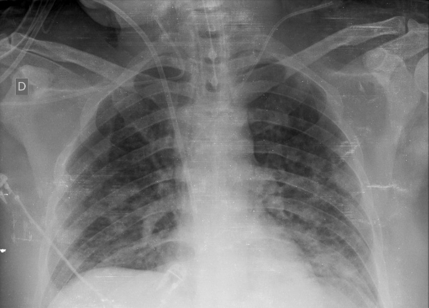
[…] to support medical decision making in imaging. However, inadequate availability of a diverse annotated dataset has limited the performance and generalizability of existing models. Purpose To create the first multi-institutional, multi-national expert annotated COVID-19 imaging dataset made freely available to the machine learning community as a research and educational resource for COVID-19 chest imaging. The Radiological Society of North America (RSNA) assembled the RSNA International COVID-19 Open Radiology Database (RICORD) collection of COVID-related imaging datasets and expert annotations to support research and education. RICORD data will be incorporated in the Medical Imaging and Data Resource Center (MIDRC), a multi-institutional research data repository funded by the National Institute of Biomedical Imaging and Bioengineering of the National Institutes of Health. Materials and Methods This dataset was created through a collaboration between the RSNA and Society of Thoracic Radiology (STR). Clinical annotation by thoracic radiology subspecialists was performed for all COVID positive chest radiography (CXR) imaging studies using a labeling schema based upon guidelines for reporting classification of COVID-19 findings in CXRs (see Review of Chest Radiograph Findings of COVID-19 Pneumonia and Suggested Reporting Language, Journal of Thoracic Imaging). Results The RSNA International COVID-19 Open Annotated Radiology Database (RICORD) consists of 998 chest x-rays from 361 patients at four international sites annotated with diagnostic labels. Patient Selection: Patients at least 18 years in age receiving positive diagnosis for COVID-19. Data Abstract 998 Chest x-ray examinations from 361 patients. Annotations with labels: Classification Typical Appearance Multifocal bilateral, peripheral opacities, and/or Opacities with rounded morphology Lower lung-predominant distribution (Required Feature – must be present with either or both of the first two opacity patterns) Indeterminate Appearance Absence of typical findings AND Unilateral, central or upper lung predominant distribution of airspace disease Atypical Appearance Pneumothorax or pleural effusion, Pulmonary Edema, Lobar Consolidation, Solitary lung nodule or mass, Diffuse tiny nodules, Cavity Negative for Pneumonia No lung opacities Airspace Disease Grading Lungs are divided on frontal chest xray into 3 zones per lung (6 zones total). The upper zone extends from the apices to the superior hilum. The mid zone spans between the superior and inferior hilar margins. The lower zone extends from the inferior hilar margins to the costophrenic sulci. Mild – Required if not negative for pneumonia Opacities in 1-2 lung zones Moderate – Required if not negative for pneumonia Opacities in 3-4 lung zones Severe – Required if not negative for pneumonia Opacities in >4 lung zones Supporting clinical […]


